03 June 2024 | Monday | News

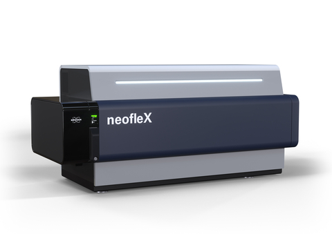
Bruker Corporation announced the launch of a novel, high-performance MALDI-TOF/TOF system, the neofleX Imaging Profiler for mass spectrometry-based tissue imaging. It enables facile OME-TIFF file output via the new SCiLS Scope software. The transformative neofleX MALDI-TOF/TOF MSI system now conveniently fits on a bench-top.
The neofleX Imaging Profiler MALDI-TOF/TOF mass spectrometer comes standard with Bruker’s proprietary 10 kHz smartbeam 3D laser for true pixel fidelity and with enhanced imaging detectors designed for longitudinal robustness, stability, and reproducibility in linear and reflector modes. neofleX is also available as a TOF/TOF configuration that features a reimagined fragmentation module for significantly improved TOF/TOF sensitivity, speed and sequence coverage.
Created for the unmet needs of moving from discovery imaging to translational and clinical tissue research, neofleX was used by the group of Prof. Bernd Bodenmiller at ETH and University of Zuerich to simultaneously map 116 proteins across a lung FFPE tissue section in 7 hours, using the MALDI HiPLEX-IHC workflow. Multiplexed detection with neofleX and MALDI HiPLEX-IHC technology allows increasing the number of proteins to map cellular processes without increasing MSI measurement time for a given region of interest.
In addition to MALDI HiPLEX-IHC MSI immunohistochemistry, neofleX is also compatible with the MALDI-ISH (in situhybridization) method announced at ASMS 2024 by AmberGen, Inc. MALDI-ISH multiplexes imaging of up to 12 oligomers of interest (RNA/DNA) for multiomic spatial tissue research in neuroscience, infectious disease, and oncology.
The novel neofleX excels at providing more insight per pixel through multiomic spatial biology data from tissue sections that can positively correlate targeted proteins with glycans, metabolites, lipids, endogenous peptides, xenobiotics, and now RNA/DNA. This additional multiomics context provides important adjacency information about cellular states, function, structure, and protein activity for a range of research areas, such as oncology and neurology.
Professor Carsten Hopf of the University of Mannheim, Germany, Center for Mass Spectrometry and Optical Spectroscopy (CeMOS) commented: “The innovative neofleX presents an incredibly powerful, versatile and easy-to-use mass-spec imaging system for tissue spatial biology researchers for targeted proteomics. We appreciate the value of this instrument for performance and versatility, and our clinical research collaborators welcome the translational research capabilities.”
Multiomic co-localization of lipids and glycans on a tissue section allows to not only localize protein targets using MALDI HiPLEX-IHC, but also assess protein activity and function. A study performed at CeMOS on brain tissues from a transgenic mouse model demonstrated co-localization of amyloid β-42 (Aβ42) protein with a targeted membrane-bound glycosphingolipid (GM3 d36:1), yielding important structural information.
Dr. Michael L. Easterling, Vice President MSI for Bruker’s Life Sciences Mass Spectrometry division, commented: “The neofleX offers unique combination of outstanding performance, multimodal software and workflows on the benchtop for a wide range of biopharma and clinical researchers. Additionally, neofleX brings novel capabilities to spatial biology, including spatial proteomics combined with unique multiomics correlations for developing actionable biological insights.”
The neofleX is compatible with Bruker’s MALDI Imaging software and consumables ecosystems, such as IntelliSlides™ and SCiLS autopilot that simplify sample tracking, preparation, and analysis and require minimal input from users to initiate and process automated mass-spec imaging runs. For ease of collaborations, the neofleX now delivers targeted imaging data via automatically generated OME-TIFF images that can be viewed within the SCiLS environment, or easily exported into custom pipelines or digital pathology systems.
Bruker also announced extension of the SCiLS portfolio with SCiLS Scope 1.0 for collaboration around targeted, multiomic translational workflows developed for neofleX. SCiLS Scope software supports OME-TIFF datasets from targeted imaging workflows such as MALDI HiPLEX-IHC. Ion images are visualized by false-color coding of selected channels, and image processing and distance measurements can be accomplished with simple tools.
Blaine R. Roberts, Ph.D., Associate Professor in the Department of Biochemistry and Department of Neurology at Emory University in Atlanta, GA, summarized: “Fast and user-friendly visualization of targets is made easy by the addition of SCiLS Scope to the MSI software lineup.”
© 2026 Biopharma Boardroom. All Rights Reserved.