16 May 2023 | Tuesday | News

By PerkinElmer
Non-invasive approach
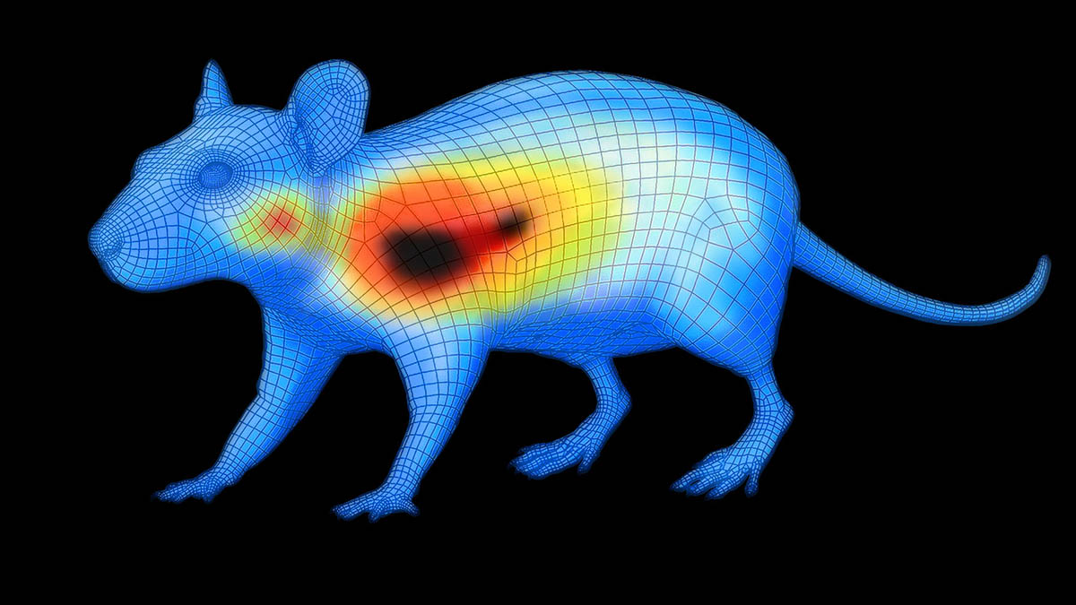
In vivo fluorescence imaging is a non-invasive technique that uses light to visualize biological processes. The ability to image the same animals longitudinally reduces the number of animals required for experiments.
Early information
Imaging performed at the end of a study will likely miss key biological processes. In vivo fluorescence imaging provides opportunities to monitor changes in biology prior to overt physical changes. It can also detect therapeutic response shortly after treatment (prior to surrogate or clinical endpoints) and characterize multiple biomarkers over the course of disease (multiplex imaging).
Broad applications
In oncology research, in vivo fluorescence imaging can be used to predict changes in tumor burden, growth, regression, and metastasis based on early, quantifiable changes in proteases, cell surface receptors, or vascularity. Beyond oncology, fluorescence imaging can be utilized to study complex biology and detect early biological changes, biomarkers, and mechanisms that underlie other diseases such as liver and kidney toxicity, arthritis, or autoimmunity.
Functionality and versatility
Importantly, fluorescence imaging can be used for tracking CAR-NK cells or other cells that resist bioluminescent labeling, or when a luciferase causes an immune response. Fluorescent probes can also provide correlative data when used in combination with bioluminescence, microCT, or PET imaging for multimodal studies.
A key aspect of successful in vivo imaging is to set clear goals for your study and identify which imaging approach will provide the most appropriate benefits. For example, you need to decide whether your primary goal is to explore early or late disease and then decide which imaging approach will draw the most useful conclusions for your investigation.
Successful fluorescence imaging requires carefully chosen probes that have been validated for your biology of interest. Additionally, probes with high specificity and biologically appropriate characteristics, such as clearance rate and route of metabolism, will enable unique and meaningful biological information to be extracted from your studies.
At PerkinElmer, our fluorescent imaging probes are developed through an extensive R&D process, designed to incorporate drug-like biodistribution properties, and validated for optimal target delivery and performance. Each probe’s structure, size, specificity, and physiochemical properties are optimized to achieve the ideal pharmacokinetics, biodistribution, and metabolic clearance characteristics essential for interrogating the relevant biology.
To help inform your decisions and advance your oncology research, we have developed an interactive Selector Guide for IVISense™ fluorescent probes. By matching probe properties to specific biology and biomarker characteristics, you can use the guide to better understand how imaging and quantification of early biological changes associated with disease development, therapeutic efficacy, and drug-induced tissue changes can be achieved.

© 2026 Biopharma Boardroom. All Rights Reserved.